Hands-On Breast Ultrasound Imaging and Doppler
A one-day guided experience that prepares you to detect subtle change with precision, judgment, and trust at the bedside.
“I feel confident performing a quality breast ultrasound.”
-Eleanor Glass
Get Here. Learn It. Practice It. Serve.
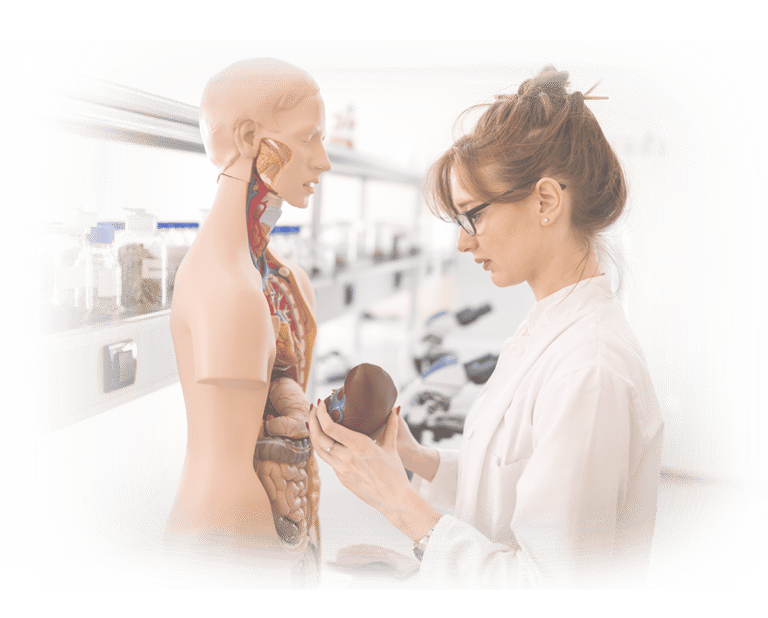
When Pattern Matters.
On this day, hands-on skills will start to become second nature, not second guess. This course meets you exactly where you are and takes you exactly where you need to go. From the moment you begin, you’ll feel the difference: a calm, welcoming environment, expert instructors at your side, and a step-by-step approach that removes the intimidation and replaces it with clarity, purpose, and confidence.
Every concept is explained in plain, practical language—anatomy, physics, instrumentation, Doppler, and pathology—so you always understand why you’re doing something before we show you how to do it. Then, nearly every minute is devoted to hands-on scanning with real-time, individual guidance that builds a repeatable breast imaging workflow you can trust in any clinical setting.
You'll leave with a clear protocol, stronger visual reasoning, and the confidence to evaluate breast anatomy with precision and care. And because your growth doesn’t stop at the end of the day, your post-course support continues forever—free of charge, whenever you need us. Here, you’re never on your own.
Command the Invisible for the Good of the Patient—Why This Course Matters For You.
Breast imaging demands precision with sensitivity—clinical and interpersonal. This hands-on course matters because it teaches you to quietly detect subtle change and discern between benign and concerning patterns in a structured, guided setting.
You’ll refine image acquisition, Doppler assessment, and interpretation strategies that allow you to make confident clinical calls and communicate them effectively to patients and care teams.
What Makes This Course Different.
You won't just watch someone scan — you'll spend most of the entire day scanning live patients with step-by-step coaching, simple explanations, and supportive mentoring that meets you where you are. You’ll leave with a foundation strong enough to build a career of clinical service on— service in compassionate care to the hundreds of patients you've yet to meet.
How Each Professional Benefits by Attending.
Radiologic Technologists
Integrates mammography with ultrasound skills so you can correlate findings with confidence and elevate their diagnostic contribution
Improves lesion characterization skills, enabling you to distinguish cystic vs. solid structures quickly and accurately.
Builds mastery of targeted scanning protocols, giving them the ability to support radiologists more effectively and expand their clinical role
Creates a career-advancing competency that many breast centers now prefer—or require—in technologists.
Improves lesion characterization skills, enabling you to distinguish cystic vs. solid structures quickly and accurately.
General Sonographers
Provides a repeatable, anatomy-anchored workflow that removes the guesswork and anxiety of breast orientation
Strengthens understanding of quadrant and radial scanning, Doppler applications, and documentation standards
Bridges the gap between general abdominal/MSK scanning and the highly specific demands of breast imaging
Nurse Practitioners / PA's.
Enables clinicians to perform focused, point-of-care breast evaluations right in the exam room to speed decision-making
Enhances their ability to triage palpable findings, guide next steps, and communicate more precisely with radiology
Helps them develop a systematic, defensible documentation method that reduces uncertainty and strengthens clinical confidence
Adds a high-impact skill that improves patient trust, continuity of care, and practice efficiency..
Newer Clinicians.
Provides a repeatable, anatomy-anchored workflow that removes the guesswork and anxiety of breast orientation
Introduces and strengthens understanding of quadrant and radial scanning, Doppler applications, and documentation standards
Bridges the gap between general abdominal scanning and the highly specific demands of breast imaging
Offers immediate, hands-on repetition so their scanning confidence rises in real time.
Medical Device Professionals. Research, Engineering, Marketing, Sales, Management, Applications, Service Engineers
Deep Clinical Understanding. Experience the exact challenges clinicians face at the bedside — fuel better product design, training and messaging.
Stronger Conversations & Credibility. Speak the clinician’s language. Demonstrate relevance, credibility, and insight in customer discussions — no guesswork.
No prior breast scanning experience is required—only your desire and commitment to learn.
What You'll Learn Clinically.
A comprehensive step-by-step breast ultrasound protocol you can use immediately in clinical practice
All lecture is woven into your practice with live scanning all day with expert guidance—no observation-only learning
Master a tactile technique proven to increase lesion detection sensitivity by nearly 15%
Build confidence in both quadrant and radial scanning methods for complete breast coverage
Understand the “why” behind image and Doppler findings with easy-to-grasp physics explanations
Learn how to document and communicate normal vs. abnormal findings with accuracy and clarity
Gain lifetime access to post-class mentoring from instructors with 40+ years of real-world experience
A skillset that makes you more valuable in breast imaging, women’s health, and diagnostic triage.
There’s no test, no pressure—just guided practice, real feedback, and hands-on skill building.
Our Approach.
Ultrasound skill is built through guided experience, not passive observation. Instruction centers on live scanning, immediate feedback, and repeated practice—so technique, interpretation, and clinical reasoning develop together.
We teach in small groups to allow faculty to adjust instruction in real time, responding to each learner’s background, pace, and questions. Concepts are revisited from multiple angles, helping understanding take root quickly and remain reliable under clinical pressure.
The emphasis is not on memorizing images or following rigid protocols, but on learning how to think at the bedside—recognizing patterns, understanding physiology, and making deliberate decisions that translate directly to patient care.
What Our Participants Said.
The overwhelming majority described the experience as excellent, transformational, and more effective for them than other national ultrasound courses.
Students repeatedly mention they learned more in a few days than in months elsewhere.
Course Details at a Glance.
Format: Live, in-person, hands-on instruction.
Length: One day: 9am - 4pm.
Location: Las Colinas→(City of Irving, Dallas–Fort Worth area).
Class Size: Intentionally limited to four attendees for scan time and faculty access.
Scan Lab: Open access for extended practice.
Instruction Style: Small-group, guided scanning with immediate feedback.
Tuition—Your Career Investment.
Smart Career Growth— Zero Debt→
This investment reflects the micro-class size, intensity of hands-on instruction, detailed course materials you'll reference forever, unlimited scan-lab access, and lifetime post-course mentoring. Hot breakfast and light lunch are included daily.
Tuition: $1000. Saturday, 9am-3pm.
If tuition is a concern, we’ve already thought about that.
Download this guided Employer-Sponsorship Agreement Kit→ that lets your workplace pay for the course while you agree to stay for a defined time afterward. It’s one of the easiest ways to advance your skills without taking on new expenses.
Most Attendees say the Course pays for itself in just a few weeks of bedside application.
About CME Credit.
This course does not offer CME credit—by design.
That choice allows us to teach with complete flexibility, adapting instruction to your background, pace, and clinical goals rather than a committee's preset agenda.
Our focus is hands-on skill development, clinical reasoning, and lasting bedside confidence. Many clinicians choose to pair this experience with separate CME activities that align with their own professional requirements. If we can help direct you to such low- or no-cost CEM resources, we'lll be delighted to assist.


You've Got Questions?
Let's face them together.
Your decision is transformational. Ask us anything and get a straight answer. We want to address your every question, because You and what you’re about to do are important.

Your Next Step Is Closer—and Brighter—Than You Think.
You've come so far already— don't quit.
You’ve considered the most demanding parts: the time apart from the people who matter, the disruptions of travel, the honest moments of doubt, and the uncertainty we all feel when something truly matters. Here, you are safe. Here, you are supported. And what you gain will elevate you, your family, and your patients for years to come.
It would be easier—and much more hollow—to simply turn on the TV or computer and watch a video. But every lasting accomplishment comes by doing. You don’t learn ultrasound skills sitting on your hands.
Start your success now—without hesitation:
Download your free Anatomy Charts and Reference Guides→
Connect directly with Keith→ on LinkedIn for personal guidance
Request to join our private Alumni Community→ on Facebook.
Your Journey is already underway.
We’ll meet you in the Scan Lab soon—and at the Bedside for the rest of your career....
Training Clinicians Worldwide Since 1981.
Testimonials reflect individual learning experiences. Growth in skill and confidence develops through guided training, continued practice, and personal commitment.
Contact Us
(972 | 353-3200 USA Central Time
Course Campus:
4300 Wingren Drive
Irving (Las Colinas), Texas 75039
Mail | FedEx Address:
Box 101
Colleyville, TX 76034
Legal & Administrative
Privacy Policy
Accessibility Statement
© Keith Mauney & Associates Ultrasound Training Institutes MMXXVI. All rights reserved.
Courses & Learning
Your Skills-Building Experience
Hands-On POCUS- Point of Care Ultrasound
Hands-On Adult Echocardiography
Hands-On Transvaginal Pelvic Ultrasound
Ultrasound Physics
Cardiovascular Hemodynamics
EKG- From Atoms to Arrhythmias