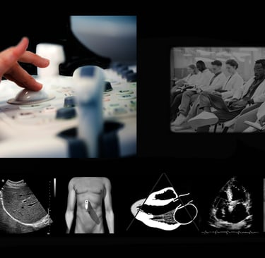
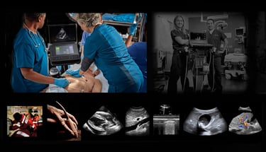
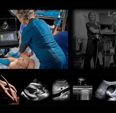
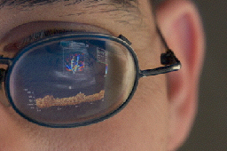
From Probe to Practice.
Real-life ultrasound learning moments and expert guidance.
Introduction to Ultrasound for Clinicians New to It Est. 1984
This beginner-level, fully hands-on Course brings you into the scan lab from the very start, teaching you how to hold the probe, orient the image, understand anatomy in motion, and begin applying ultrasound as a practical clinical tool.
You'll appreciate that in this course:
You’ll finally get to learn ultrasound with your hand on the transducer—not just watching someone else do it.
Small-group scanning means you get direct, continuous coaching from expert clinicians who know how to teach, not just perform.
Every concept is taught at the bedside, in real time, so anatomy, physics, and image optimization all make sense together, not in isolation.
You’ll gain confidence quickly by practicing on real anatomy, real equipment, and real cases—not simulations or prerecorded video.
You’ll leave with a clear, repeatable protocol you can use immediately in practice—plus a 100+ page reference manual that takes home every step, image, and even a simple, practical walkthrough of ultrasound physics and instrumentation.


Monday - Friday
Hands-On POCUS Ultrasound Course Est. 1992
Originally created for the trauma surgery service at Parkland Memorial Hospital in Dallas, we've extracted every key element needed for your bedside or on-scene ultrasound success. These support the FAST, RUSH and FATE protocols used worldwide today. You'll get clear, step-by-step guidance in a small-group setting so you can quickly feel confident using ultrasound at the bedside or in the field in everyday patient encounters..
You'll appreciate that this course:
Focuses on practical skills you can use immediately, not abstract theory.
Lets you learn by doing, with expert support at your side—not from slides or lectures.
Builds confidence through repetition, real-time feedback, and guided scanning on live models.
Meets you at your current skill level, whether you’re brand-new or refining what you already know.
Breaks down the pedantry of physics and instrumentation into digestible bites to address your future POCUS Academy certification.
Continues to support you after class with your own comprehensive manual, and direct access to instructors for questions and follow-up help.

Friday - Saturday
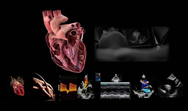
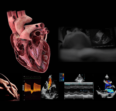
Hands-On Cardiac Ultrasound Imaging & Doppler Adult Echocardiography Course Est. 1976
We'll carefully escort you through a comprehensive, streamlined, real-world protocol—explaining the “why” behind every view, every Doppler survey, and every measurement, so you can deliver exactly what the cardiologist needs with skill and professional authority.
You'll appreciate that this course is built around:
A truly simplified, real-world protocol that finally makes echo feel logical, repeatable, and clinically useful—not overwhelming.
Hands-on scanning in the very first hours, with expert mentors at your shoulder—not a lecture, not a demo, but real practice with real equipment.
Respect for your role as a clinician—your questions are welcomed, your experience is honored, and your time isn’t wasted with theory that never reaches the bedside.
Practical pearls, shortcuts, and “street-smart” tips that only come from decades in the field—not found in textbooks or slide decks... and everything that's covered (and more) goes home with you in your 200 page manual.
Monday - Friday
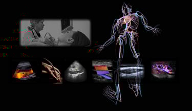
Hands-On Vascular Ultrasound Arterial & Venous Imaging & Doppler Course Est. 1978
We'll build with you the mental and tactile tools to perform vascular ultrasound with authority—capturing the images, Doppler data, and interpretive markers that surgeons, vascular medicine specialists, and interventional teams actually depend on.
You'll appreciate how this Course focuses on You:
You’ll finally see vascular ultrasound organized into a clear, step-by-step structure—making sense of what is normally taught as a maze of unrelated tests.
Instead of memorizing anatomy and waveforms, you’ll learn the logic behind blood flow, pressure gradients, and hemodynamics—so every image and Doppler signal has purpose.
Hands-on scanning replaces passive watching, giving you repeated practice on real anatomy with expert guidance at your shoulder, not across a lecture hall.
Complex arterial and venous exams are broken down into repeatable bedside protocols you can use the day you return in the clinic, lab, or OR.- and they're all documented in your own 125-page reference manual.
You’ll build confidence not only in what to record, but how to interpret it—understanding what the vascular surgeon, cardiologist, or interventionalist is actually looking for.
Monday - Thursday

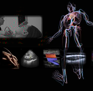
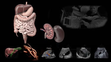
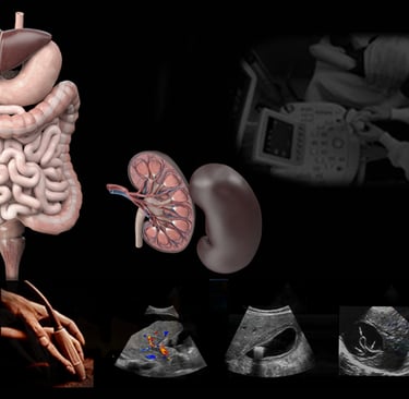
Hands-On Abdominal Ultrasound Imaging & Doppler Course Est. 1989
We'll equip you with the skills, speed, and clinical reasoning to perform abdominal studies with confidence—so the images you produce answer questions, not create more of them. With your hands on the probe, we'll show you exactly what to scan, what to document, and how to make every image clinically meaningful. You’ll leave with a complete, proven protocol for abdominal imaging—plus the lifelong mentoring to keep growing long after class ends.
You'll appreciate how this Course focuses on You:
You’ll finally learn abdominal ultrasound the way it makes sense—organ by organ, with a clear bedside protocol instead of disconnected images and trivia.
Hands-on scanning starts right away, so you’re not just watching someone else scan—you’re learning by doing, with expert guidance at your shoulder.
You’ll understand why each view, measurement, and Doppler sweep matters clinically, not just how to get the image.
Complex anatomy becomes manageable when you see it on real patients in real time, instead of only in textbooks or static diagrams.
You’ll leave knowing how to produce a complete, defensible abdominal study that answers real diagnostic questions—not just fills in worksheet blanks... and your own 130-page reference manual will serve you for life..

Monday - Wednesday
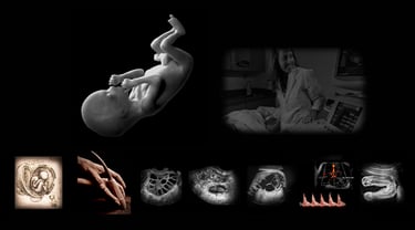
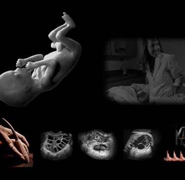
Hands-On Obstetric Ultrasound Imaging & Doppler Course Est. 1989
Together, we're going to carefully move through a rigorously structured, scan-intensive experience that sharpens your tactile skill, spatial reasoning, and diagnostic judgment—so you can perform a complete fetal anatomy and Doppler study with clarity, precision, and lasting confidence.
You'll appreciate these five learning advantages:
A fully repeatable cervix-to-fundus protocol that ensures every fetal structure is located, imaged, measured, and documented with scientific consistency—not guesswork.
Deliberate practice on live pregnant subjects from day one, reinforced by immediate faculty coaching, peer repetition, and evening open-lab access for self-directed scanning on yourself or class peers.
Deep integration of spectral, color, and power Doppler—not as an afterthought, but as a core element of decision-making for fetal growth, placental integrity, and high-risk pregnancy assessment.
Expert instruction rooted in 40+ years of ultrasound education and clinical consulting, designed to collapse the normal three-year learning curve into four days of guided immersion.
Permanent post-course support and career guidance, including protocol reinforcement, case troubleshooting, credentialing strategy, and direct access to faculty long after you leave the classroom.
Monday - Thursday
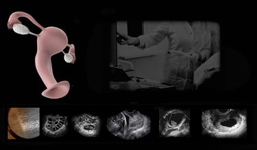
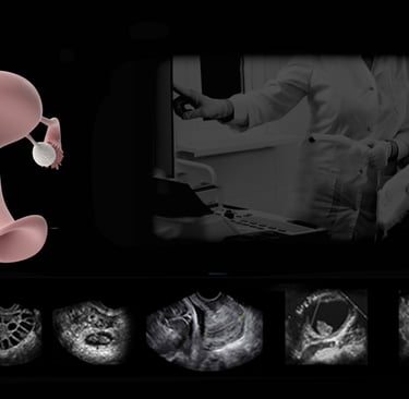
Hands-On Transvaginal Pelvic Ultrasound Imaging & Doppler Course Est. 1989
This one-day course gives you an intensive, protocol-driven, scan-focused experience that equips you to confidently identify, document, and analyze every pelvic structure—with the Doppler mastery, spatial orientation skills, and post-training support needed for true independent clinical practice.
You'll appreciate how this Course focuses on:
A precise, repeatable pelvic protocol that ensures you can always locate the uterus, adnexa, ovaries, bladder, pelvic floor, and pouch of Douglas—without losing orientation or second-guessing anatomy.
Hands-on scanning from the start, reinforced by direct faculty coaching, so your hand-eye coordination and anatomic recognition accelerate in days—not months.
Clear integration of color, power, and spectral Doppler with standardized settings and interpretation strategies for vascularity, perfusion, and mass characterization.
Instruction in real-world problem-solving techniques including artifact recognition, ovary-verification logic, adnexal pathology differentiation, and when to shift to translabial or surface pelvic imaging.
A small-group format with unrestricted Q&A and case-based discussion, ideal for clinicians returning to practice, expanding scope, or preparing for credentialing and independent scanning.
Free lifetime post-course mentoring, giving you direct access to expert support as you apply protocols, troubleshoot images, and grow into full clinical autonomy.

Friday
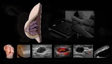
Hands-On Breast Ultrasound Imaging & Doppler Course Est. 1983
If you’ve ever wished breast ultrasound felt clearer, more systematic, and more clinically intuitive, this hands-on course was built for you. Together, we’ll walk through every layer of breast anatomy—from the skin surface to the chest wall—and show you how to translate what you already know from training into a sharper, more confident approach at the bedside. Nearly the entire day is devoted to guided scanning on live volunteers, giving you the chance to truly feel the workflow, refine your image optimization, and see the real differences between normal tissue, benign findings, and suspicious features. Nothing is rushed, and nothing important is skipped—whether physics, protocol, or pathology. By the end of the day, you’ll have a clean, repeatable method you can rely on in every exam.
You'll appreciate these six learning advantages:
Supportive hands-on scanning with live volunteers, so you’re not just watching—you’re practicing, correcting, and building real clinical confidence.
A simple, repeatable protocol that helps you stay oriented and consistent, no matter which patient, machine, or breast type you’re imaging.
Clear explanation of benign vs. malignant patterns, with the “why” behind each feature so you’re not just memorizing appearances—you’re understanding them.
Practical Doppler instruction that shows you what vascular patterns should look like and how they support your interpretation.
Training on universal system controls, helping you finally master the adjustments that make the biggest difference in image quality.
Ongoing access to us for questions and support, so you’re never left wondering whether you’re doing it right once you’re back at work.
Saturday

Frequently Asked
Who can attend courses?
Healthcare professionals of any specialty or experience level can join our hands-on ultrasound courses. There are no prerequisites, but we encourage you to discuss
Where are courses held?
Courses take place in Dallas, Texas, or conveniently at your own clinical site for on-site training.
What makes training unique?
How we teach is what sets us apart.
We prioritize hands-on practice, explain the why behind every step, and translate abstract science into clear clinical meaning. Visit our Attendee Voices page to hear it directly from those who’ve trained with us.
Is mentoring included?
Few educational experiences extend beyond the classroom—we believe they should.
Free ongoing mentoring is included for life, with direct access to our Program Director to support real clinical questions, whenever they arise.
Because learning ultrasound is a career, not an event.

Training Clinicians Worldwide Since 1981.
Testimonials reflect individual learning experiences. Growth in skill and confidence develops through guided training, continued practice, and personal commitment.
Contact Us
(972 | 353-3200 USA Central Time
Course Campus:
4300 Wingren Drive
Irving (Las Colinas), Texas 75039
Mail | FedEx Address:
Box 101
Colleyville, TX 76034
Legal & Administrative
Privacy Policy
Accessibility Statement
© Keith Mauney & Associates Ultrasound Training Institutes MMXXVI. All rights reserved.
Courses & Learning
Your Skills-Building Experience
Hands-On POCUS- Point of Care Ultrasound
Hands-On Adult Echocardiography
Hands-On Transvaginal Pelvic Ultrasound
Ultrasound Physics
Cardiovascular Hemodynamics
EKG- From Atoms to Arrhythmias