How the Hands On Cardiac Ultrasound class typically unfolds.
Our cardiac course unfolds the same way true understanding develops—step by step, never rushed. And tailored to your learning style.
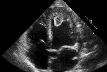
We begin by establishing a shared visual language: cardiac anatomy as it appears on the screen, with careful attention to orientation, probe position, and spatial relationships.
Scanning starts early. Learners rotate through short, focused scan sessions with faculty at the bedside, repeating views and refining technique until confidence builds naturally.
Once images stabilize, physiology is layered in. Wall motion, valve function, timing, and Doppler are introduced only after the image makes sense.
Concepts are revisited throughout the course so understanding deepens, not resets. Small groups allow faculty to adapt teaching in real time.
The scan lab remains open beyond scheduled hours, giving learners freedom to practice and reinforce skills.
By the end of the course, learners leave with a clear, repeatable approach to cardiac scanning—and continued mentoring long after the class adjourns.

Training Clinicians Worldwide Since 1981.
Testimonials reflect individual learning experiences. Growth in skill and confidence develops through guided training, continued practice, and personal commitment.
Contact Us
(972 | 353-3200 USA Central Time
Course Campus:
4300 Wingren Drive
Irving (Las Colinas), Texas 75039
Mail | FedEx Address:
Box 101
Colleyville, TX 76034
Legal & Administrative
Privacy Policy
Accessibility Statement
© Keith Mauney & Associates Ultrasound Training Institutes MMXXVI. All rights reserved.
Courses & Learning
Your Skills-Building Experience
Hands-On POCUS- Point of Care Ultrasound
Hands-On Adult Echocardiography
Hands-On Transvaginal Pelvic Ultrasound
Ultrasound Physics
Cardiovascular Hemodynamics
EKG- From Atoms to Arrhythmias