After the Course—From Class to Bedside
A few anchors to carry forward as your experience continues to unfold.
You’ve just completed an intense, hands-on experience—one designed not to end at the classroom door, but to keep working quietly in the background as you move forward. What follows is a recap of how we approached your training, why we did it that way, and where you can continue to draw support as your experience deepens.
Congratulations! Here's a list of the resources we discussed in Class.
Our 7-Step Approach to Your Training:
Vision.
In a brief time, we’ve put the technical and clinical pictures together in the context of separate diagnosis and management pathways… all in the context of the global patient and provider care setting, to grow sharper with every case, forever. It's been to equip you further for your own Journey.
Stay with us in Linkedin→ and Born to Scan→ to keep the conversation close.
Vocabulary.
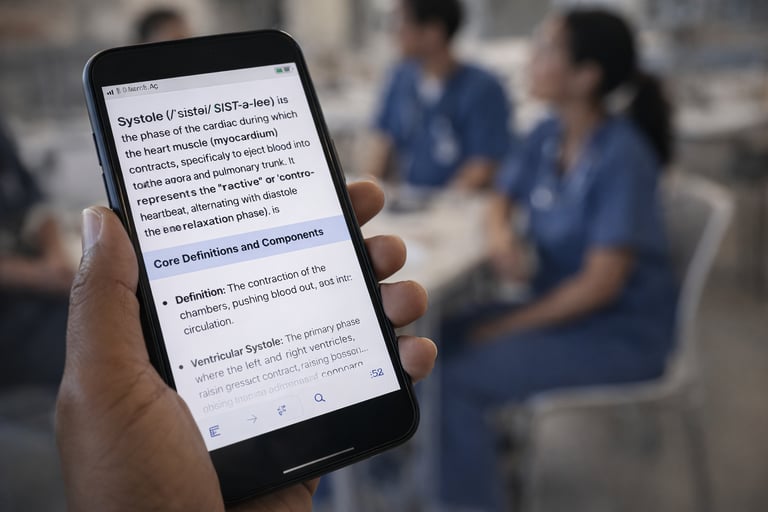
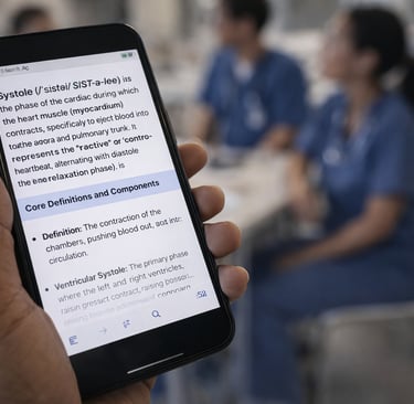
As you recall, we looked behind the face of every pertinent term to find where it came from, as its specific meaning is important, even though the different specialties have hijacked them for their own field. We matched them to their in-the-street-language so you, too, can pass them on clearly and more permanently concrete their precise meaning and value.
Download the free Merriam-Webster Dictionary→ app.
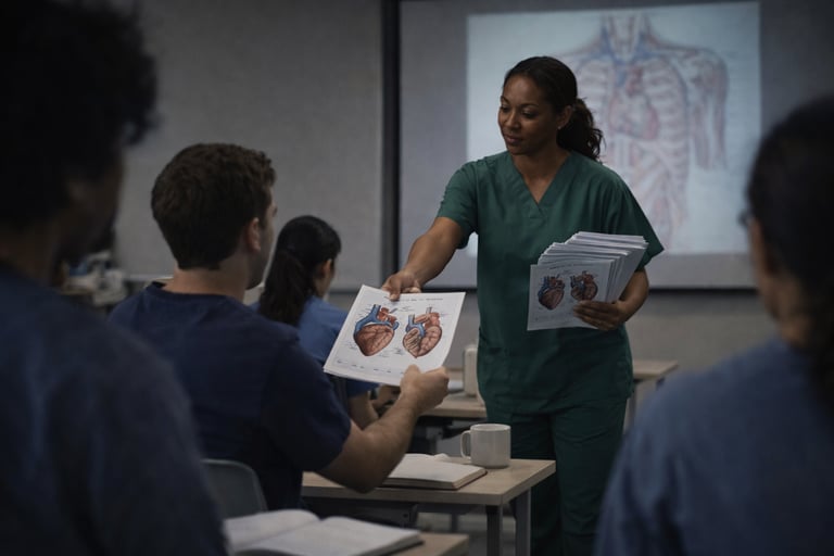
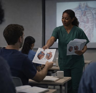
Anatomy.
Download these anatomy charts→ to your cell phone; also print & paste them everywhere in sight. You’re visual; make the most of it.
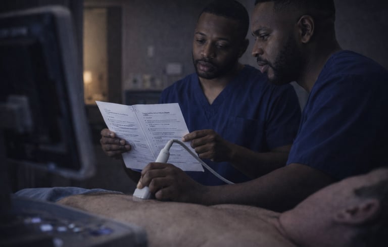
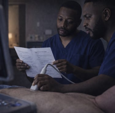
Protocol.
Remember? It started with the fingertip probe grasp (with the fifth finger down, to ensure you’ll gain a year-and-a-half jump start. And we set in motion, we simplified, and canonized the universal foundation and process on which you and your team will forever build.
Practice.
You’ll never get enough; that’s why we’ll be standing beside you in perpetuity to help share our collective thinking together, for free. It's just not how many cases you've done; it's how you think with each one you do.
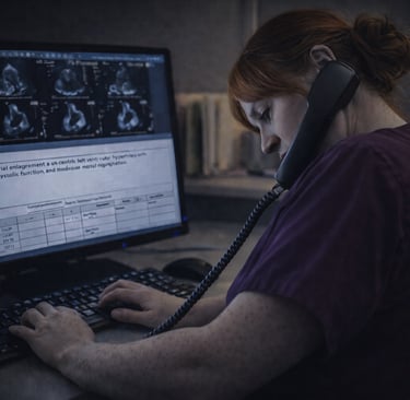
Pathology.
Like every important responsible professional, you need ready-references at your fingertips:
POCUS: ThePOCUSatlas.com→
Abdominal, Vascular: Ultrasoundcases.info→
Abdominal/OB/Gyn: Ultrasound-Images.com→
Cardiac: Echocardiographer.org→ Echo Atlas→

Communication.
If we did our job in Class properly, we said everything at least three different ways by at least two different means, all pitched to our best guess of what words and timing would be best for you. Now it’s time for all those apparently disparate facts, analogies and stories to unwind, ferment, and reveal over time their career-long intent for You. And now it's time for that line of communication to reach out into your very far and successful Future.. Because, now, everyone is going to want to hear what you have to say.
We've built our approach around your career in Service. And we're still here for you. Call on us→ for anything, any time. The answer is already Yes.
Training Clinicians Worldwide Since 1981.
Testimonials reflect individual learning experiences. Growth in skill and confidence develops through guided training, continued practice, and personal commitment.
Contact
972 | 353-3200 USA Central Time
Course Campus:
4300 Wingren Drive
Irving (Las Colinas), Texas 75039
Mail | FedEx Address:
Box 101
Colleyville, TX 76034
Courses & Learning
Ultrasound Physics
Cardiovascular Hemodynamics
EKG- From Atoms to Arrhythmias